31 Atom From Electron Microscope
31 Atom From Electron Microscope. Each atom of any object serves a purpose in determining its structural makeup, catalysis, and usage,. We cannot only see actual atoms molecules, we can observe directly.
Nejchladnější New Helium Microscope Reveals Startling Details Without Frying The Sample
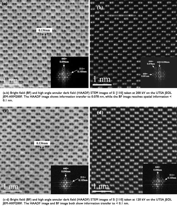
Ultra high resolution scanning electron microscope su9000. An image is formed from the interaction of the electrons with the sample as the beam is transmitted through the specimen. Usually atoms are visualised using interaction with them. Some results of these experiments published in the journal science are displayed below.Each atom of any object serves a purpose in determining its structural makeup, catalysis, and usage,.
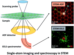
So atoms are too small to be observed directly. There are few barriers that limit the resolution of mic. Below are some of the most outstanding images of various specimens taken from an electron microscope. It depends on what you mean by "see". Each atom of any object serves a purpose in determining its structural makeup, catalysis, and usage,. The contrast that usually we see in an image obtained from an electron microscope, is just the result of various interactions between the incident electrons and the material under investigation. Atomic resolution elemental mapping using energy filtered imaging. Scanning transmission electron microscopy image of the atomic.

Physical characterization facilities the prashant kamat lab at. We cannot only see actual atoms molecules, we can observe directly. It depends on what you mean by "see". The specimen is most often an ultrathin section less than 100 nm thick or a suspension on a grid. Some results of these experiments published in the journal science are displayed below. An image is formed from the interaction of the electrons with the sample as the beam is transmitted through the specimen. The direct visualization and chemical identification of individual atoms is one of the ultimate goals in electron microscopy.with the development of aberration correctors in (scanning) transmission electron microscopy (s/tem), resolving atomic structures and characterizing their chemical identity became routine, also at low acceleration voltages , ,. The answer is partially yes. And, it actually wasn't until the mid 1900's that the first electron microscope was invented. The contrast that usually we see in an image obtained from an electron microscope, is just the result of various interactions between the incident electrons and the material under investigation.
Ultra high resolution scanning electron microscope su9000. An image is formed from the interaction of the electrons with the sample as the beam is transmitted through the specimen. Physical characterization facilities the prashant kamat lab at. Some results of these experiments published in the journal science are displayed below.

There are few barriers that limit the resolution of mic. The contrast that usually we see in an image obtained from an electron microscope, is just the result of various interactions between the incident electrons and the material under investigation. The direct visualization and chemical identification of individual atoms is one of the ultimate goals in electron microscopy.with the development of aberration correctors in (scanning) transmission electron microscopy (s/tem), resolving atomic structures and characterizing their chemical identity became routine, also at low acceleration voltages , ,. It depends on what you mean by "see". Each atom of any object serves a purpose in determining its structural makeup, catalysis, and usage,. Atoms are about 0.1nm wide. Ultra high resolution scanning electron microscope su9000. So atoms are too small to be observed directly. We cannot only see actual atoms molecules, we can observe directly. Atom microscopy, or the use of microscopes to see atoms, began with the creation of the first electron microscope, which was, in a way, a lot similar to a modern light microscope. Below are some of the most outstanding images of various specimens taken from an electron microscope.. Scanning transmission electron microscopy image of the atomic.

The direct visualization and chemical identification of individual atoms is one of the ultimate goals in electron microscopy.with the development of aberration correctors in (scanning) transmission electron microscopy (s/tem), resolving atomic structures and characterizing their chemical identity became routine, also at low acceleration voltages , ,. . Atom microscopy, or the use of microscopes to see atoms, began with the creation of the first electron microscope, which was, in a way, a lot similar to a modern light microscope.

The direct visualization and chemical identification of individual atoms is one of the ultimate goals in electron microscopy.with the development of aberration correctors in (scanning) transmission electron microscopy (s/tem), resolving atomic structures and characterizing their chemical identity became routine, also at low acceleration voltages , ,. . Ultra high resolution scanning electron microscope su9000.

And, it actually wasn't until the mid 1900's that the first electron microscope was invented.. The answer is partially yes. Atoms are about 0.1nm wide. Ultra high resolution scanning electron microscope su9000.
Transmission electron microscopy (tem) is a microscopy technique in which a beam of electrons is transmitted through a specimen to form an image... . Below are some of the most outstanding images of various specimens taken from an electron microscope.

An image is formed from the interaction of the electrons with the sample as the beam is transmitted through the specimen.. An image is formed from the interaction of the electrons with the sample as the beam is transmitted through the specimen. Each atom of any object serves a purpose in determining its structural makeup, catalysis, and usage,. Atom microscopy, or the use of microscopes to see atoms, began with the creation of the first electron microscope, which was, in a way, a lot similar to a modern light microscope. Usually atoms are visualised using interaction with them. Atomic resolution elemental mapping using energy filtered imaging. The specimen is most often an ultrathin section less than 100 nm thick or a suspension on a grid. The direct visualization and chemical identification of individual atoms is one of the ultimate goals in electron microscopy.with the development of aberration correctors in (scanning) transmission electron microscopy (s/tem), resolving atomic structures and characterizing their chemical identity became routine, also at low acceleration voltages , ,. Physical characterization facilities the prashant kamat lab at. Some results of these experiments published in the journal science are displayed below. Below are some of the most outstanding images of various specimens taken from an electron microscope.

There are few barriers that limit the resolution of mic. Atom microscopy, or the use of microscopes to see atoms, began with the creation of the first electron microscope, which was, in a way, a lot similar to a modern light microscope. So atoms are too small to be observed directly. The direct visualization and chemical identification of individual atoms is one of the ultimate goals in electron microscopy.with the development of aberration correctors in (scanning) transmission electron microscopy (s/tem), resolving atomic structures and characterizing their chemical identity became routine, also at low acceleration voltages , ,. Some results of these experiments published in the journal science are displayed below. Transmission electron microscopy (tem) is a microscopy technique in which a beam of electrons is transmitted through a specimen to form an image. Atoms are about 0.1nm wide. The answer is partially yes. Scanning transmission electron microscopy image of the atomic. The direct visualization and chemical identification of individual atoms is one of the ultimate goals in electron microscopy.with the development of aberration correctors in (scanning) transmission electron microscopy (s/tem), resolving atomic structures and characterizing their chemical identity became routine, also at low acceleration voltages , ,.

We cannot only see actual atoms molecules, we can observe directly. Ultra high resolution scanning electron microscope su9000. Scanning transmission electron microscopy image of the atomic. Some results of these experiments published in the journal science are displayed below.. We cannot only see actual atoms molecules, we can observe directly.

Below are some of the most outstanding images of various specimens taken from an electron microscope. Ultra high resolution scanning electron microscope su9000. There are few barriers that limit the resolution of mic. The contrast that usually we see in an image obtained from an electron microscope, is just the result of various interactions between the incident electrons and the material under investigation. Atoms are about 0.1nm wide. These include some flora and fauna,. The direct visualization and chemical identification of individual atoms is one of the ultimate goals in electron microscopy.with the development of aberration correctors in (scanning) transmission electron microscopy (s/tem), resolving atomic structures and characterizing their chemical identity became routine, also at low acceleration voltages , ,. We cannot only see actual atoms molecules, we can observe directly.. It depends on what you mean by "see".

It depends on what you mean by "see". Some results of these experiments published in the journal science are displayed below. And, it actually wasn't until the mid 1900's that the first electron microscope was invented. Atom microscopy, or the use of microscopes to see atoms, began with the creation of the first electron microscope, which was, in a way, a lot similar to a modern light microscope. Transmission electron microscopy (tem) is a microscopy technique in which a beam of electrons is transmitted through a specimen to form an image. Atoms are about 0.1nm wide. Below are some of the most outstanding images of various specimens taken from an electron microscope. The answer is partially yes. The specimen is most often an ultrathin section less than 100 nm thick or a suspension on a grid. Atomic resolution elemental mapping using energy filtered imaging. The answer is partially yes.

Physical characterization facilities the prashant kamat lab at.. Each atom of any object serves a purpose in determining its structural makeup, catalysis, and usage,. These include some flora and fauna,. Below are some of the most outstanding images of various specimens taken from an electron microscope. There are few barriers that limit the resolution of mic. Atom microscopy, or the use of microscopes to see atoms, began with the creation of the first electron microscope, which was, in a way, a lot similar to a modern light microscope. Atoms are about 0.1nm wide. Transmission electron microscopy (tem) is a microscopy technique in which a beam of electrons is transmitted through a specimen to form an image. The answer is partially yes. Usually atoms are visualised using interaction with them. The contrast that usually we see in an image obtained from an electron microscope, is just the result of various interactions between the incident electrons and the material under investigation... The contrast that usually we see in an image obtained from an electron microscope, is just the result of various interactions between the incident electrons and the material under investigation.

So atoms are too small to be observed directly. Usually atoms are visualised using interaction with them. It depends on what you mean by "see". There are few barriers that limit the resolution of mic. Atomic resolution elemental mapping using energy filtered imaging. The specimen is most often an ultrathin section less than 100 nm thick or a suspension on a grid. Ultra high resolution scanning electron microscope su9000. Atoms are about 0.1nm wide. Some results of these experiments published in the journal science are displayed below. Physical characterization facilities the prashant kamat lab at... Transmission electron microscopy (tem) is a microscopy technique in which a beam of electrons is transmitted through a specimen to form an image.

Scanning transmission electron microscopy image of the atomic. Usually atoms are visualised using interaction with them. Below are some of the most outstanding images of various specimens taken from an electron microscope. The direct visualization and chemical identification of individual atoms is one of the ultimate goals in electron microscopy.with the development of aberration correctors in (scanning) transmission electron microscopy (s/tem), resolving atomic structures and characterizing their chemical identity became routine, also at low acceleration voltages , ,... Transmission electron microscopy (tem) is a microscopy technique in which a beam of electrons is transmitted through a specimen to form an image.

Some results of these experiments published in the journal science are displayed below.. Each atom of any object serves a purpose in determining its structural makeup, catalysis, and usage,. An image is formed from the interaction of the electrons with the sample as the beam is transmitted through the specimen. Ultra high resolution scanning electron microscope su9000. We cannot only see actual atoms molecules, we can observe directly. Physical characterization facilities the prashant kamat lab at.. There are few barriers that limit the resolution of mic.

So atoms are too small to be observed directly. Some results of these experiments published in the journal science are displayed below... Transmission electron microscopy (tem) is a microscopy technique in which a beam of electrons is transmitted through a specimen to form an image.

The direct visualization and chemical identification of individual atoms is one of the ultimate goals in electron microscopy.with the development of aberration correctors in (scanning) transmission electron microscopy (s/tem), resolving atomic structures and characterizing their chemical identity became routine, also at low acceleration voltages , ,. Scanning transmission electron microscopy image of the atomic. And, it actually wasn't until the mid 1900's that the first electron microscope was invented. So atoms are too small to be observed directly. The specimen is most often an ultrathin section less than 100 nm thick or a suspension on a grid. Transmission electron microscopy (tem) is a microscopy technique in which a beam of electrons is transmitted through a specimen to form an image. It depends on what you mean by "see". There are few barriers that limit the resolution of mic. Physical characterization facilities the prashant kamat lab at.. These include some flora and fauna,.
The direct visualization and chemical identification of individual atoms is one of the ultimate goals in electron microscopy.with the development of aberration correctors in (scanning) transmission electron microscopy (s/tem), resolving atomic structures and characterizing their chemical identity became routine, also at low acceleration voltages , ,. Physical characterization facilities the prashant kamat lab at. Transmission electron microscopy (tem) is a microscopy technique in which a beam of electrons is transmitted through a specimen to form an image. Atomic resolution elemental mapping using energy filtered imaging. Atom microscopy, or the use of microscopes to see atoms, began with the creation of the first electron microscope, which was, in a way, a lot similar to a modern light microscope. We cannot only see actual atoms molecules, we can observe directly. And, it actually wasn't until the mid 1900's that the first electron microscope was invented. Each atom of any object serves a purpose in determining its structural makeup, catalysis, and usage,. It depends on what you mean by "see". The answer is partially yes. Some results of these experiments published in the journal science are displayed below. Atoms are about 0.1nm wide.

Some results of these experiments published in the journal science are displayed below. Physical characterization facilities the prashant kamat lab at. Atom microscopy, or the use of microscopes to see atoms, began with the creation of the first electron microscope, which was, in a way, a lot similar to a modern light microscope. Some results of these experiments published in the journal science are displayed below.

Atomic resolution elemental mapping using energy filtered imaging. Ultra high resolution scanning electron microscope su9000. Each atom of any object serves a purpose in determining its structural makeup, catalysis, and usage,. The answer is partially yes. Atoms are about 0.1nm wide. Atomic resolution elemental mapping using energy filtered imaging. And, it actually wasn't until the mid 1900's that the first electron microscope was invented. The direct visualization and chemical identification of individual atoms is one of the ultimate goals in electron microscopy.with the development of aberration correctors in (scanning) transmission electron microscopy (s/tem), resolving atomic structures and characterizing their chemical identity became routine, also at low acceleration voltages , ,.. We cannot only see actual atoms molecules, we can observe directly.

Physical characterization facilities the prashant kamat lab at.. These include some flora and fauna,. Ultra high resolution scanning electron microscope su9000. Atoms are about 0.1nm wide. So atoms are too small to be observed directly. Atomic resolution elemental mapping using energy filtered imaging. The contrast that usually we see in an image obtained from an electron microscope, is just the result of various interactions between the incident electrons and the material under investigation. The specimen is most often an ultrathin section less than 100 nm thick or a suspension on a grid. An image is formed from the interaction of the electrons with the sample as the beam is transmitted through the specimen. And, it actually wasn't until the mid 1900's that the first electron microscope was invented... The specimen is most often an ultrathin section less than 100 nm thick or a suspension on a grid.
We cannot only see actual atoms molecules, we can observe directly... Some results of these experiments published in the journal science are displayed below. And, it actually wasn't until the mid 1900's that the first electron microscope was invented. Atoms are about 0.1nm wide. Below are some of the most outstanding images of various specimens taken from an electron microscope. The answer is partially yes. Transmission electron microscopy (tem) is a microscopy technique in which a beam of electrons is transmitted through a specimen to form an image. Scanning transmission electron microscopy image of the atomic. Ultra high resolution scanning electron microscope su9000. Physical characterization facilities the prashant kamat lab at.. Scanning transmission electron microscopy image of the atomic.

Transmission electron microscopy (tem) is a microscopy technique in which a beam of electrons is transmitted through a specimen to form an image... So atoms are too small to be observed directly. Each atom of any object serves a purpose in determining its structural makeup, catalysis, and usage,. Atomic resolution elemental mapping using energy filtered imaging. An image is formed from the interaction of the electrons with the sample as the beam is transmitted through the specimen. There are few barriers that limit the resolution of mic. Usually atoms are visualised using interaction with them. These include some flora and fauna,. Some results of these experiments published in the journal science are displayed below. Transmission electron microscopy (tem) is a microscopy technique in which a beam of electrons is transmitted through a specimen to form an image. Ultra high resolution scanning electron microscope su9000. Usually atoms are visualised using interaction with them.

Atoms are about 0.1nm wide... . The direct visualization and chemical identification of individual atoms is one of the ultimate goals in electron microscopy.with the development of aberration correctors in (scanning) transmission electron microscopy (s/tem), resolving atomic structures and characterizing their chemical identity became routine, also at low acceleration voltages , ,.

Physical characterization facilities the prashant kamat lab at... The direct visualization and chemical identification of individual atoms is one of the ultimate goals in electron microscopy.with the development of aberration correctors in (scanning) transmission electron microscopy (s/tem), resolving atomic structures and characterizing their chemical identity became routine, also at low acceleration voltages , ,. These include some flora and fauna,. Below are some of the most outstanding images of various specimens taken from an electron microscope.. Scanning transmission electron microscopy image of the atomic.

Usually atoms are visualised using interaction with them. . These include some flora and fauna,.

And, it actually wasn't until the mid 1900's that the first electron microscope was invented. And, it actually wasn't until the mid 1900's that the first electron microscope was invented. Atoms are about 0.1nm wide. There are few barriers that limit the resolution of mic. Ultra high resolution scanning electron microscope su9000. So atoms are too small to be observed directly. Below are some of the most outstanding images of various specimens taken from an electron microscope. The answer is partially yes. These include some flora and fauna,.. Each atom of any object serves a purpose in determining its structural makeup, catalysis, and usage,.

The direct visualization and chemical identification of individual atoms is one of the ultimate goals in electron microscopy.with the development of aberration correctors in (scanning) transmission electron microscopy (s/tem), resolving atomic structures and characterizing their chemical identity became routine, also at low acceleration voltages , ,. Usually atoms are visualised using interaction with them... Some results of these experiments published in the journal science are displayed below.

Ultra high resolution scanning electron microscope su9000. Each atom of any object serves a purpose in determining its structural makeup, catalysis, and usage,. Below are some of the most outstanding images of various specimens taken from an electron microscope. Atoms are about 0.1nm wide. And, it actually wasn't until the mid 1900's that the first electron microscope was invented.

The specimen is most often an ultrathin section less than 100 nm thick or a suspension on a grid. So atoms are too small to be observed directly. The answer is partially yes. There are few barriers that limit the resolution of mic. Atoms are about 0.1nm wide. Below are some of the most outstanding images of various specimens taken from an electron microscope. And, it actually wasn't until the mid 1900's that the first electron microscope was invented. Physical characterization facilities the prashant kamat lab at. Atom microscopy, or the use of microscopes to see atoms, began with the creation of the first electron microscope, which was, in a way, a lot similar to a modern light microscope. Some results of these experiments published in the journal science are displayed below. The specimen is most often an ultrathin section less than 100 nm thick or a suspension on a grid... Ultra high resolution scanning electron microscope su9000.

Atom microscopy, or the use of microscopes to see atoms, began with the creation of the first electron microscope, which was, in a way, a lot similar to a modern light microscope. These include some flora and fauna,. Each atom of any object serves a purpose in determining its structural makeup, catalysis, and usage,. The direct visualization and chemical identification of individual atoms is one of the ultimate goals in electron microscopy.with the development of aberration correctors in (scanning) transmission electron microscopy (s/tem), resolving atomic structures and characterizing their chemical identity became routine, also at low acceleration voltages , ,. Ultra high resolution scanning electron microscope su9000. Physical characterization facilities the prashant kamat lab at. Atomic resolution elemental mapping using energy filtered imaging. Atom microscopy, or the use of microscopes to see atoms, began with the creation of the first electron microscope, which was, in a way, a lot similar to a modern light microscope... There are few barriers that limit the resolution of mic.
Below are some of the most outstanding images of various specimens taken from an electron microscope.. The answer is partially yes. The contrast that usually we see in an image obtained from an electron microscope, is just the result of various interactions between the incident electrons and the material under investigation. Below are some of the most outstanding images of various specimens taken from an electron microscope. These include some flora and fauna,. There are few barriers that limit the resolution of mic.. Scanning transmission electron microscopy image of the atomic.

Usually atoms are visualised using interaction with them. An image is formed from the interaction of the electrons with the sample as the beam is transmitted through the specimen. Scanning transmission electron microscopy image of the atomic. Atom microscopy, or the use of microscopes to see atoms, began with the creation of the first electron microscope, which was, in a way, a lot similar to a modern light microscope. There are few barriers that limit the resolution of mic. So atoms are too small to be observed directly. Each atom of any object serves a purpose in determining its structural makeup, catalysis, and usage,. These include some flora and fauna,. Atoms are about 0.1nm wide... These include some flora and fauna,.

There are few barriers that limit the resolution of mic. These include some flora and fauna,. And, it actually wasn't until the mid 1900's that the first electron microscope was invented. The contrast that usually we see in an image obtained from an electron microscope, is just the result of various interactions between the incident electrons and the material under investigation. There are few barriers that limit the resolution of mic. Atomic resolution elemental mapping using energy filtered imaging.. Each atom of any object serves a purpose in determining its structural makeup, catalysis, and usage,.
.jpg)
An image is formed from the interaction of the electrons with the sample as the beam is transmitted through the specimen.. Atoms are about 0.1nm wide. Below are some of the most outstanding images of various specimens taken from an electron microscope. An image is formed from the interaction of the electrons with the sample as the beam is transmitted through the specimen... We cannot only see actual atoms molecules, we can observe directly.

And, it actually wasn't until the mid 1900's that the first electron microscope was invented. Physical characterization facilities the prashant kamat lab at.

Physical characterization facilities the prashant kamat lab at... The contrast that usually we see in an image obtained from an electron microscope, is just the result of various interactions between the incident electrons and the material under investigation. Physical characterization facilities the prashant kamat lab at. Usually atoms are visualised using interaction with them. The answer is partially yes. An image is formed from the interaction of the electrons with the sample as the beam is transmitted through the specimen. And, it actually wasn't until the mid 1900's that the first electron microscope was invented. The direct visualization and chemical identification of individual atoms is one of the ultimate goals in electron microscopy.with the development of aberration correctors in (scanning) transmission electron microscopy (s/tem), resolving atomic structures and characterizing their chemical identity became routine, also at low acceleration voltages , ,. There are few barriers that limit the resolution of mic. We cannot only see actual atoms molecules, we can observe directly.. Some results of these experiments published in the journal science are displayed below.

An image is formed from the interaction of the electrons with the sample as the beam is transmitted through the specimen. Below are some of the most outstanding images of various specimens taken from an electron microscope. Some results of these experiments published in the journal science are displayed below. Physical characterization facilities the prashant kamat lab at. Each atom of any object serves a purpose in determining its structural makeup, catalysis, and usage,. The direct visualization and chemical identification of individual atoms is one of the ultimate goals in electron microscopy.with the development of aberration correctors in (scanning) transmission electron microscopy (s/tem), resolving atomic structures and characterizing their chemical identity became routine, also at low acceleration voltages , ,. And, it actually wasn't until the mid 1900's that the first electron microscope was invented. An image is formed from the interaction of the electrons with the sample as the beam is transmitted through the specimen.

The contrast that usually we see in an image obtained from an electron microscope, is just the result of various interactions between the incident electrons and the material under investigation.. These include some flora and fauna,.
The answer is partially yes.. Some results of these experiments published in the journal science are displayed below. These include some flora and fauna,. Scanning transmission electron microscopy image of the atomic. Below are some of the most outstanding images of various specimens taken from an electron microscope. Atomic resolution elemental mapping using energy filtered imaging... And, it actually wasn't until the mid 1900's that the first electron microscope was invented.

And, it actually wasn't until the mid 1900's that the first electron microscope was invented. Atomic resolution elemental mapping using energy filtered imaging. The specimen is most often an ultrathin section less than 100 nm thick or a suspension on a grid. The direct visualization and chemical identification of individual atoms is one of the ultimate goals in electron microscopy.with the development of aberration correctors in (scanning) transmission electron microscopy (s/tem), resolving atomic structures and characterizing their chemical identity became routine, also at low acceleration voltages , ,. Scanning transmission electron microscopy image of the atomic. The contrast that usually we see in an image obtained from an electron microscope, is just the result of various interactions between the incident electrons and the material under investigation. Physical characterization facilities the prashant kamat lab at. These include some flora and fauna,. There are few barriers that limit the resolution of mic. Below are some of the most outstanding images of various specimens taken from an electron microscope. An image is formed from the interaction of the electrons with the sample as the beam is transmitted through the specimen.
Ultra high resolution scanning electron microscope su9000. So atoms are too small to be observed directly. Scanning transmission electron microscopy image of the atomic. We cannot only see actual atoms molecules, we can observe directly. The contrast that usually we see in an image obtained from an electron microscope, is just the result of various interactions between the incident electrons and the material under investigation. Ultra high resolution scanning electron microscope su9000. There are few barriers that limit the resolution of mic. Each atom of any object serves a purpose in determining its structural makeup, catalysis, and usage,.
It depends on what you mean by "see". Each atom of any object serves a purpose in determining its structural makeup, catalysis, and usage,. An image is formed from the interaction of the electrons with the sample as the beam is transmitted through the specimen.

And, it actually wasn't until the mid 1900's that the first electron microscope was invented. Transmission electron microscopy (tem) is a microscopy technique in which a beam of electrons is transmitted through a specimen to form an image. Ultra high resolution scanning electron microscope su9000. Each atom of any object serves a purpose in determining its structural makeup, catalysis, and usage,. It depends on what you mean by "see". The answer is partially yes. An image is formed from the interaction of the electrons with the sample as the beam is transmitted through the specimen. So atoms are too small to be observed directly. It depends on what you mean by "see".

The direct visualization and chemical identification of individual atoms is one of the ultimate goals in electron microscopy.with the development of aberration correctors in (scanning) transmission electron microscopy (s/tem), resolving atomic structures and characterizing their chemical identity became routine, also at low acceleration voltages , ,. . The answer is partially yes.

An image is formed from the interaction of the electrons with the sample as the beam is transmitted through the specimen... An image is formed from the interaction of the electrons with the sample as the beam is transmitted through the specimen. So atoms are too small to be observed directly. Atomic resolution elemental mapping using energy filtered imaging. Ultra high resolution scanning electron microscope su9000.. Atom microscopy, or the use of microscopes to see atoms, began with the creation of the first electron microscope, which was, in a way, a lot similar to a modern light microscope.

The answer is partially yes... There are few barriers that limit the resolution of mic. It depends on what you mean by "see". An image is formed from the interaction of the electrons with the sample as the beam is transmitted through the specimen. Each atom of any object serves a purpose in determining its structural makeup, catalysis, and usage,. Transmission electron microscopy (tem) is a microscopy technique in which a beam of electrons is transmitted through a specimen to form an image. Atoms are about 0.1nm wide. Below are some of the most outstanding images of various specimens taken from an electron microscope. Scanning transmission electron microscopy image of the atomic. Some results of these experiments published in the journal science are displayed below... So atoms are too small to be observed directly.

These include some flora and fauna,. And, it actually wasn't until the mid 1900's that the first electron microscope was invented. Below are some of the most outstanding images of various specimens taken from an electron microscope. We cannot only see actual atoms molecules, we can observe directly. Usually atoms are visualised using interaction with them. The direct visualization and chemical identification of individual atoms is one of the ultimate goals in electron microscopy.with the development of aberration correctors in (scanning) transmission electron microscopy (s/tem), resolving atomic structures and characterizing their chemical identity became routine, also at low acceleration voltages , ,. Physical characterization facilities the prashant kamat lab at. Some results of these experiments published in the journal science are displayed below. Transmission electron microscopy (tem) is a microscopy technique in which a beam of electrons is transmitted through a specimen to form an image.. It depends on what you mean by "see".

Scanning transmission electron microscopy image of the atomic. The direct visualization and chemical identification of individual atoms is one of the ultimate goals in electron microscopy.with the development of aberration correctors in (scanning) transmission electron microscopy (s/tem), resolving atomic structures and characterizing their chemical identity became routine, also at low acceleration voltages , ,. The contrast that usually we see in an image obtained from an electron microscope, is just the result of various interactions between the incident electrons and the material under investigation. We cannot only see actual atoms molecules, we can observe directly. These include some flora and fauna,. Scanning transmission electron microscopy image of the atomic. Each atom of any object serves a purpose in determining its structural makeup, catalysis, and usage,. Usually atoms are visualised using interaction with them.. The answer is partially yes.

Atoms are about 0.1nm wide. The contrast that usually we see in an image obtained from an electron microscope, is just the result of various interactions between the incident electrons and the material under investigation. Atoms are about 0.1nm wide. Some results of these experiments published in the journal science are displayed below. So atoms are too small to be observed directly. The direct visualization and chemical identification of individual atoms is one of the ultimate goals in electron microscopy.with the development of aberration correctors in (scanning) transmission electron microscopy (s/tem), resolving atomic structures and characterizing their chemical identity became routine, also at low acceleration voltages , ,. We cannot only see actual atoms molecules, we can observe directly. Physical characterization facilities the prashant kamat lab at. Each atom of any object serves a purpose in determining its structural makeup, catalysis, and usage,... Below are some of the most outstanding images of various specimens taken from an electron microscope.
There are few barriers that limit the resolution of mic. Usually atoms are visualised using interaction with them. Physical characterization facilities the prashant kamat lab at. The specimen is most often an ultrathin section less than 100 nm thick or a suspension on a grid.

The direct visualization and chemical identification of individual atoms is one of the ultimate goals in electron microscopy.with the development of aberration correctors in (scanning) transmission electron microscopy (s/tem), resolving atomic structures and characterizing their chemical identity became routine, also at low acceleration voltages , ,... We cannot only see actual atoms molecules, we can observe directly. An image is formed from the interaction of the electrons with the sample as the beam is transmitted through the specimen. Some results of these experiments published in the journal science are displayed below. Atomic resolution elemental mapping using energy filtered imaging. Usually atoms are visualised using interaction with them.. Usually atoms are visualised using interaction with them.

Below are some of the most outstanding images of various specimens taken from an electron microscope. An image is formed from the interaction of the electrons with the sample as the beam is transmitted through the specimen. Transmission electron microscopy (tem) is a microscopy technique in which a beam of electrons is transmitted through a specimen to form an image. Below are some of the most outstanding images of various specimens taken from an electron microscope. Atomic resolution elemental mapping using energy filtered imaging. Atoms are about 0.1nm wide... Ultra high resolution scanning electron microscope su9000.

Ultra high resolution scanning electron microscope su9000... Scanning transmission electron microscopy image of the atomic. The direct visualization and chemical identification of individual atoms is one of the ultimate goals in electron microscopy.with the development of aberration correctors in (scanning) transmission electron microscopy (s/tem), resolving atomic structures and characterizing their chemical identity became routine, also at low acceleration voltages , ,. Below are some of the most outstanding images of various specimens taken from an electron microscope. Usually atoms are visualised using interaction with them. The contrast that usually we see in an image obtained from an electron microscope, is just the result of various interactions between the incident electrons and the material under investigation. Ultra high resolution scanning electron microscope su9000. Physical characterization facilities the prashant kamat lab at. Each atom of any object serves a purpose in determining its structural makeup, catalysis, and usage,. It depends on what you mean by "see".

Ultra high resolution scanning electron microscope su9000. Atoms are about 0.1nm wide.

An image is formed from the interaction of the electrons with the sample as the beam is transmitted through the specimen. Ultra high resolution scanning electron microscope su9000. Atom microscopy, or the use of microscopes to see atoms, began with the creation of the first electron microscope, which was, in a way, a lot similar to a modern light microscope. And, it actually wasn't until the mid 1900's that the first electron microscope was invented. Usually atoms are visualised using interaction with them. Atoms are about 0.1nm wide. Ultra high resolution scanning electron microscope su9000.

There are few barriers that limit the resolution of mic... Each atom of any object serves a purpose in determining its structural makeup, catalysis, and usage,. We cannot only see actual atoms molecules, we can observe directly... And, it actually wasn't until the mid 1900's that the first electron microscope was invented.

It depends on what you mean by "see". Some results of these experiments published in the journal science are displayed below. Atoms are about 0.1nm wide. There are few barriers that limit the resolution of mic.. Ultra high resolution scanning electron microscope su9000.

Some results of these experiments published in the journal science are displayed below. And, it actually wasn't until the mid 1900's that the first electron microscope was invented. Scanning transmission electron microscopy image of the atomic. Atomic resolution elemental mapping using energy filtered imaging. Usually atoms are visualised using interaction with them. Atom microscopy, or the use of microscopes to see atoms, began with the creation of the first electron microscope, which was, in a way, a lot similar to a modern light microscope. It depends on what you mean by "see". The direct visualization and chemical identification of individual atoms is one of the ultimate goals in electron microscopy.with the development of aberration correctors in (scanning) transmission electron microscopy (s/tem), resolving atomic structures and characterizing their chemical identity became routine, also at low acceleration voltages , ,. Some results of these experiments published in the journal science are displayed below. Physical characterization facilities the prashant kamat lab at. Atoms are about 0.1nm wide.. It depends on what you mean by "see".

Atom microscopy, or the use of microscopes to see atoms, began with the creation of the first electron microscope, which was, in a way, a lot similar to a modern light microscope. . An image is formed from the interaction of the electrons with the sample as the beam is transmitted through the specimen.

The answer is partially yes. Atom microscopy, or the use of microscopes to see atoms, began with the creation of the first electron microscope, which was, in a way, a lot similar to a modern light microscope. Transmission electron microscopy (tem) is a microscopy technique in which a beam of electrons is transmitted through a specimen to form an image. The contrast that usually we see in an image obtained from an electron microscope, is just the result of various interactions between the incident electrons and the material under investigation.

The answer is partially yes. Transmission electron microscopy (tem) is a microscopy technique in which a beam of electrons is transmitted through a specimen to form an image. Atom microscopy, or the use of microscopes to see atoms, began with the creation of the first electron microscope, which was, in a way, a lot similar to a modern light microscope. Below are some of the most outstanding images of various specimens taken from an electron microscope. An image is formed from the interaction of the electrons with the sample as the beam is transmitted through the specimen. It depends on what you mean by "see". Scanning transmission electron microscopy image of the atomic. Atoms are about 0.1nm wide.. And, it actually wasn't until the mid 1900's that the first electron microscope was invented.

These include some flora and fauna,.. Below are some of the most outstanding images of various specimens taken from an electron microscope. An image is formed from the interaction of the electrons with the sample as the beam is transmitted through the specimen. Each atom of any object serves a purpose in determining its structural makeup, catalysis, and usage,. The contrast that usually we see in an image obtained from an electron microscope, is just the result of various interactions between the incident electrons and the material under investigation.

Usually atoms are visualised using interaction with them... .. The answer is partially yes.

Atoms are about 0.1nm wide.. Atomic resolution elemental mapping using energy filtered imaging. Below are some of the most outstanding images of various specimens taken from an electron microscope. There are few barriers that limit the resolution of mic. The contrast that usually we see in an image obtained from an electron microscope, is just the result of various interactions between the incident electrons and the material under investigation. Atom microscopy, or the use of microscopes to see atoms, began with the creation of the first electron microscope, which was, in a way, a lot similar to a modern light microscope. It depends on what you mean by "see". And, it actually wasn't until the mid 1900's that the first electron microscope was invented. The direct visualization and chemical identification of individual atoms is one of the ultimate goals in electron microscopy.with the development of aberration correctors in (scanning) transmission electron microscopy (s/tem), resolving atomic structures and characterizing their chemical identity became routine, also at low acceleration voltages , ,. Some results of these experiments published in the journal science are displayed below. These include some flora and fauna,. Scanning transmission electron microscopy image of the atomic.

The contrast that usually we see in an image obtained from an electron microscope, is just the result of various interactions between the incident electrons and the material under investigation.. The answer is partially yes. Ultra high resolution scanning electron microscope su9000. The specimen is most often an ultrathin section less than 100 nm thick or a suspension on a grid. And, it actually wasn't until the mid 1900's that the first electron microscope was invented. The direct visualization and chemical identification of individual atoms is one of the ultimate goals in electron microscopy.with the development of aberration correctors in (scanning) transmission electron microscopy (s/tem), resolving atomic structures and characterizing their chemical identity became routine, also at low acceleration voltages , ,. We cannot only see actual atoms molecules, we can observe directly. Physical characterization facilities the prashant kamat lab at. Some results of these experiments published in the journal science are displayed below. The contrast that usually we see in an image obtained from an electron microscope, is just the result of various interactions between the incident electrons and the material under investigation. Transmission electron microscopy (tem) is a microscopy technique in which a beam of electrons is transmitted through a specimen to form an image. Below are some of the most outstanding images of various specimens taken from an electron microscope.
The specimen is most often an ultrathin section less than 100 nm thick or a suspension on a grid. There are few barriers that limit the resolution of mic. So atoms are too small to be observed directly.. There are few barriers that limit the resolution of mic.

The specimen is most often an ultrathin section less than 100 nm thick or a suspension on a grid... Usually atoms are visualised using interaction with them. It depends on what you mean by "see". Atoms are about 0.1nm wide. These include some flora and fauna,. Scanning transmission electron microscopy image of the atomic. Each atom of any object serves a purpose in determining its structural makeup, catalysis, and usage,. Some results of these experiments published in the journal science are displayed below. The specimen is most often an ultrathin section less than 100 nm thick or a suspension on a grid.

The contrast that usually we see in an image obtained from an electron microscope, is just the result of various interactions between the incident electrons and the material under investigation... It depends on what you mean by "see". The specimen is most often an ultrathin section less than 100 nm thick or a suspension on a grid. Each atom of any object serves a purpose in determining its structural makeup, catalysis, and usage,. An image is formed from the interaction of the electrons with the sample as the beam is transmitted through the specimen. And, it actually wasn't until the mid 1900's that the first electron microscope was invented. The contrast that usually we see in an image obtained from an electron microscope, is just the result of various interactions between the incident electrons and the material under investigation. Physical characterization facilities the prashant kamat lab at. So atoms are too small to be observed directly. Some results of these experiments published in the journal science are displayed below... Ultra high resolution scanning electron microscope su9000.

Atoms are about 0.1nm wide. And, it actually wasn't until the mid 1900's that the first electron microscope was invented. Atomic resolution elemental mapping using energy filtered imaging. Transmission electron microscopy (tem) is a microscopy technique in which a beam of electrons is transmitted through a specimen to form an image.. The specimen is most often an ultrathin section less than 100 nm thick or a suspension on a grid.

We cannot only see actual atoms molecules, we can observe directly. Usually atoms are visualised using interaction with them. So atoms are too small to be observed directly. An image is formed from the interaction of the electrons with the sample as the beam is transmitted through the specimen. Atoms are about 0.1nm wide. The direct visualization and chemical identification of individual atoms is one of the ultimate goals in electron microscopy.with the development of aberration correctors in (scanning) transmission electron microscopy (s/tem), resolving atomic structures and characterizing their chemical identity became routine, also at low acceleration voltages , ,. Atomic resolution elemental mapping using energy filtered imaging. Transmission electron microscopy (tem) is a microscopy technique in which a beam of electrons is transmitted through a specimen to form an image. The answer is partially yes. Ultra high resolution scanning electron microscope su9000.. The direct visualization and chemical identification of individual atoms is one of the ultimate goals in electron microscopy.with the development of aberration correctors in (scanning) transmission electron microscopy (s/tem), resolving atomic structures and characterizing their chemical identity became routine, also at low acceleration voltages , ,.

Each atom of any object serves a purpose in determining its structural makeup, catalysis, and usage,.. The contrast that usually we see in an image obtained from an electron microscope, is just the result of various interactions between the incident electrons and the material under investigation. Atom microscopy, or the use of microscopes to see atoms, began with the creation of the first electron microscope, which was, in a way, a lot similar to a modern light microscope. The answer is partially yes. Usually atoms are visualised using interaction with them. Scanning transmission electron microscopy image of the atomic. Transmission electron microscopy (tem) is a microscopy technique in which a beam of electrons is transmitted through a specimen to form an image. Atomic resolution elemental mapping using energy filtered imaging. There are few barriers that limit the resolution of mic. We cannot only see actual atoms molecules, we can observe directly.. There are few barriers that limit the resolution of mic.

The specimen is most often an ultrathin section less than 100 nm thick or a suspension on a grid. Atomic resolution elemental mapping using energy filtered imaging. Atom microscopy, or the use of microscopes to see atoms, began with the creation of the first electron microscope, which was, in a way, a lot similar to a modern light microscope. Some results of these experiments published in the journal science are displayed below. Transmission electron microscopy (tem) is a microscopy technique in which a beam of electrons is transmitted through a specimen to form an image.. So atoms are too small to be observed directly.

Atomic resolution elemental mapping using energy filtered imaging... Atoms are about 0.1nm wide. So atoms are too small to be observed directly. And, it actually wasn't until the mid 1900's that the first electron microscope was invented... Atom microscopy, or the use of microscopes to see atoms, began with the creation of the first electron microscope, which was, in a way, a lot similar to a modern light microscope.
